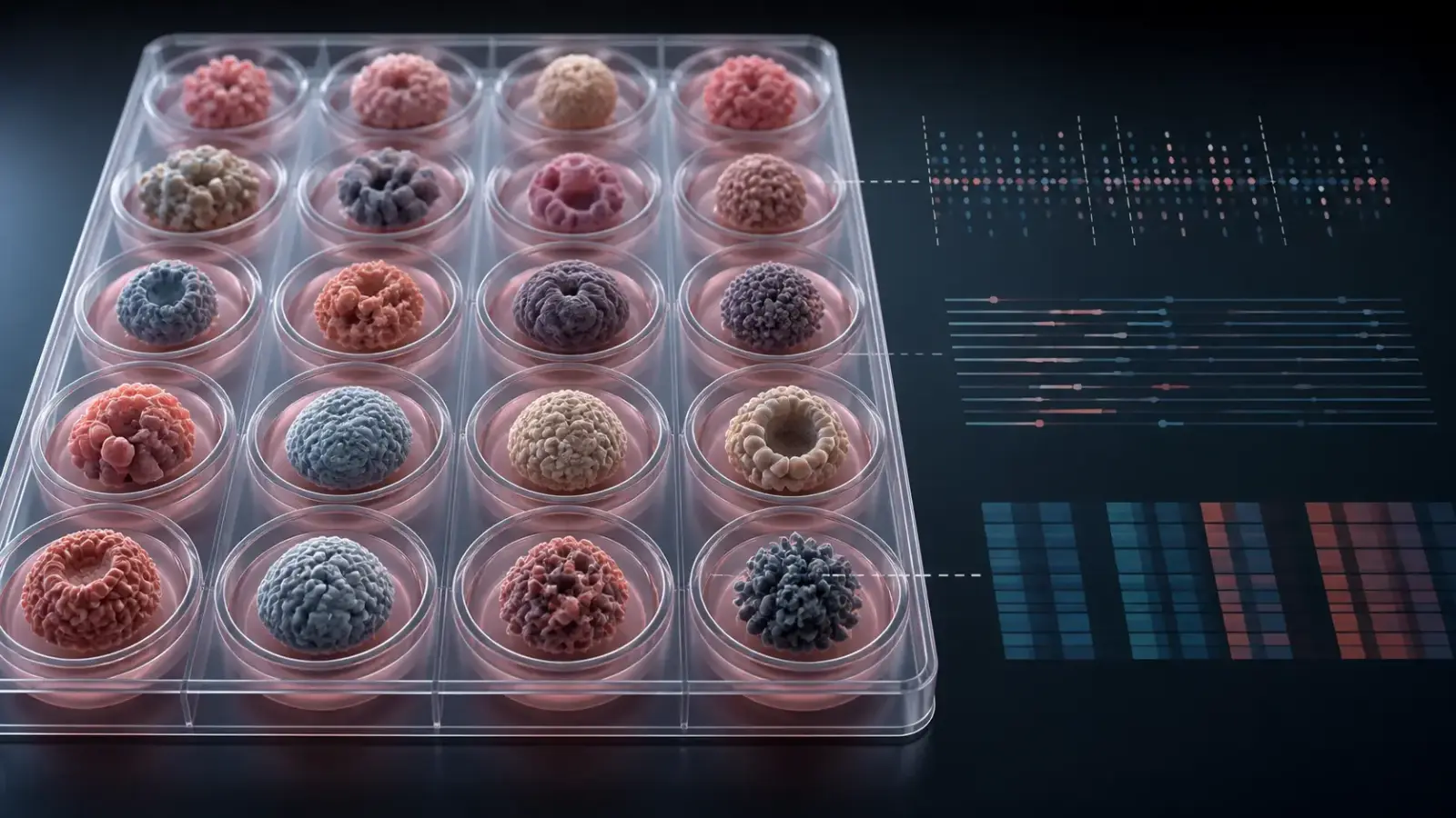
The Human Cancer Models Initiative has released 665 laboratory-grown cancer models along with something just as important: a detailed test of how closely many of them still resemble the tumors they came from.
The collection, described in Nature on August 5, spans 25 malignancies and includes three-dimensional organoids and spheroids as well as two-dimensional patient-derived cell lines. Most models preserved the major DNA, methylation and gene-expression features the researchers measured. But the study also found cases in which growth conditions favored different cell states, and it documents biological context that these cultures do not contain.
That combination makes the resource more useful than a simple claim of fidelity would. Researchers can obtain a model, inspect its relationship to the source tumor and decide whether it fits the question they want to ask.
Three numbers that answer different questions
The headline counts are easy to collapse into one another. They should not be.
According to the full study, 2,780 patients in the United States, United Kingdom, Italy and the Netherlands consented to participate between 2016 and 2021. The production and quality-control process yielded 665 models from 637 patients; some patients contributed more than one model. The researchers also examined tumor tissue from 168 failed derivation attempts.
The paper does not say that all 2,780 consented patients underwent a model-derivation attempt. Dividing 665 by 2,780 would therefore not produce a valid success rate. Establishment rates varied by cancer, disease stage, tissue type and sample quality, but the study does not report one program-wide rate with 2,780 as its denominator.
The third important count is 421: the number of models for which a matched parental tumor was available for the initial DNA comparison. Not every assay could be run on every pair. Twenty-two low-purity models were later deprioritized, leaving 643 for subsequent analyses in the paper.
What is actually in the library
Of the 665 cultures, 519 are three-dimensional organoids, 37 are three-dimensional spheroids and 109 are two-dimensional adherent cell lines. Within those broad formats are models grown by different methods, including neurospheres and conditionally reprogrammed cells. The mix matters because a growth system that works for one tumor type or experiment may be a poor fit for another.
The initiative is an international collaboration involving the National Cancer Institute, Cancer Research UK, the Wellcome Sanger Institute and Hubrecht Organoid Technology, with several model-development centers. The physical cultures were authenticated and banked for distribution through ATCC. Molecular data are available through the NCI Genomic Data Commons, and the continuously updated HCMI catalog lets researchers filter models by diagnosis, stage, treatment information, demographics, growth format and selected alterations. Some patient-associated data require controlled access rather than unrestricted download.
Clinical annotation is substantial but not universal. The paper reports comprehensive clinical data for 522 models, or 78% of the collection. It also includes 153 models from rare cancer types.
Representation remains a limitation. Genetic ancestry was estimated for 664 models, and the paper reports that 71 came from donors classified as having primarily non-European ancestry. That is about 11% of the collection. The category combines several continental ancestry groups, while some donors had complex admixture; it should not be confused with self-identified race or ethnicity. The collection broadens what is available without representing the full diversity of people or tumors affected by cancer.
“Concordance” is not one measurement
The percentages highlighted in the NIH announcement summarize several analyses, each with its own data and definition.
For DNA, the paper gives two closely related summaries. It reports that 97.8% of the assessed models retained at least two, and often all four, of the features considered: driver alterations, mutational signatures, whole-genome-doubling status and ploidy. Later, it describes 365 of 373 evaluable models as genetically concordant, rounded to 98%. The prose does not state a denominator for the 97.8% figure, so it should not be treated as a more precise restatement of 365 out of 373. Neither result means that 97.8% of every model’s mutations were identical to its tumor’s. Across the tumor-specific driver events examined, for example, 81% were retained in the corresponding models.
The 95% epigenetic result has a different denominator and test. Among 201 pairs with DNA-methylation profiles, 190 had tumor–model distances smaller than expected by chance under the study’s statistical threshold. Nine of the 11 apparently discordant pairs came from tumors estimated to contain less than 60% tumor tissue, suggesting that sample purity explained much of that signal.
RNA brought another set of definitions. One method, Celligner, classified 242 of 297 pairs, or 81%, as significantly concordant. The widely cited 92% figure comes from combining six methods: 263 of 286 models evaluated by at least four of them were flagged as divergent by no more than one method. It is a useful robustness check, not a finding that their RNA was 92% identical.
These distinctions are not statistical trivia. A model can preserve broad genomic structure while losing a specific driver, changing the abundance of a cell state or failing to retain an unstable DNA element that matters to the experiment.
Culture can purify, select and reshape
To investigate discordant cases in greater detail, the team performed single-nucleus RNA sequencing on 16 matched pairs: seven glioblastomas, six pancreatic cancers and three colorectal cancers. The analysis pointed to three different processes.
First, a culture can become “purer” in a misleading sense because stromal and immune cells present in the original tumor disappear. Second, one cancer subclone can outgrow others. Third, malignant cells can shift their transcriptional state in response to the culture environment.
The clearest medium effect appeared in glioblastoma. In one comparison, 37 models grown in a defined neural stem-cell medium were more similar to their parental tumors than three grown in conditioned medium. Switching the medium for one model changed its morphology, a stem-cell marker and gene-expression programs. The researchers did not find comparably large media effects in the other cancer lineages they assessed, so this result should not be generalized to every model and every recipe.
Extrachromosomal DNA provided a different warning. These DNA circles can carry amplified oncogenes, but their agreement between tumor and culture was far lower than the headline genomic figure: of 212 events, 43.9% were detected only in models and 32.5% only in tumors. The authors attributed much of the discordance to uneven sequencing coverage, while also noting that growth conditions can select for or against an amplification.
A tool for resistance research, not bedside prediction
The linked molecular and treatment histories make the library useful for studying why therapies stop working. The researchers identified 234 models with variants listed in a reference set of known resistance alterations. In glioblastoma, they connected prior temozolomide exposure and mismatch-repair defects with characteristic mutational signatures.
They then tested temozolomide in the laboratory. Models carrying the SBS11 signature associated with acquired resistance were less sensitive to the drug than models without it, independent of MGMT-promoter methylation in that assay. This is a concrete example of what the resource can do: connect a patient’s treatment history, a molecular trace left in the cancer and a measurable response in culture.
It is not a clinical validation study. The researchers did not prospectively assign therapy using model results or test whether those results predicted patient outcomes. The HCMI cultures also lack the stromal and immune cells needed to reproduce an intact tumor microenvironment. The paper says they may help study tumor-intrinsic mechanisms of immunotherapy resistance, not the full response to immunotherapy in a person.
The value is a better-documented stand-in
No cancer model is a miniature patient. Different questions require different features: a DNA-targeted mechanism may depend on preserving a mutation, while an immunotherapy experiment requires cellular interactions absent from these cultures. Even a highly concordant model can be wrong for a particular task.
HCMI’s advance is to make that choice more inspectable. Laboratories can work with the same authenticated material, review the same molecular reference and see which features survived culture and which did not. A surprising result is then easier to reproduce—and easier to challenge.
The library will need deeper representation and model systems that restore more of the tumor’s surroundings. Its fidelity scores are not a seal of authenticity. They are a map of where each stand-in is likely to be informative, which is ultimately more useful.