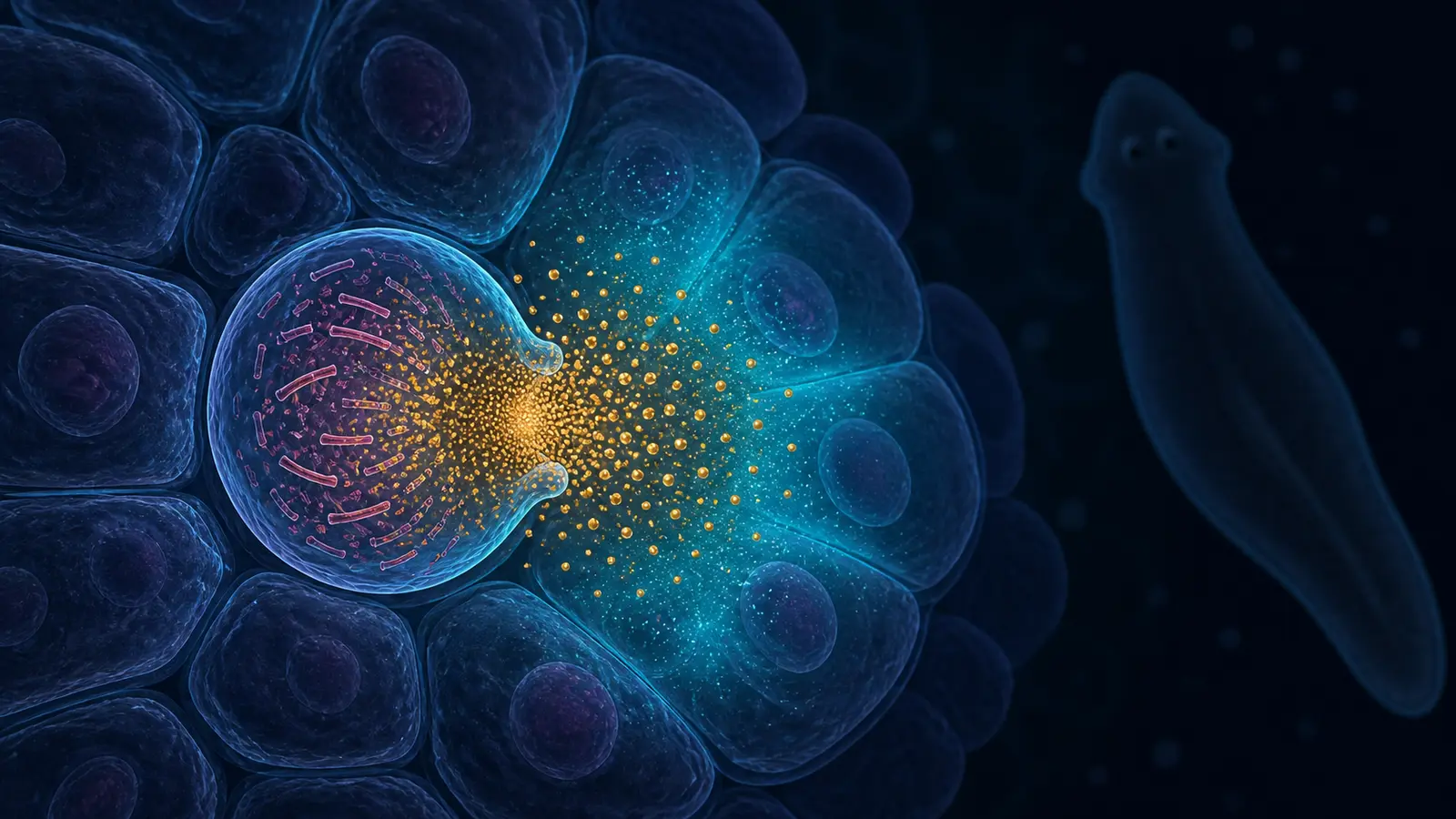
Most familiar killer cells survive the act of killing. A cytotoxic T cell or natural killer cell releases a focused molecular payload into its target and can move on. A newly described cell in planarian flatworms follows a much more terminal strategy: it disassembles its own nucleus and cytoskeleton, ruptures, and disperses a short-lived toxin into the immediate neighborhood.
The researchers who observed the event named the cell a “ruptoblast” and its death “ruptosis.” Under the microscope, the sequence can begin about a minute after stimulation and leave the original cell effectively gone within minutes.
That violence makes the discovery easy to sensationalize. Its real importance is more precise. Ruptoblasts are glandular cells rather than blood-lineage immune cells, yet experiments indicate that they help reject incompatible tissue and control bacteria. They expand the list of cell types that evolution has recruited for cytotoxic defense.
A rejection experiment led to an unexpected cell
The study began with a basic question about self and non-self in planarians. These freshwater flatworms are famous for regeneration: a population of adult stem cells called neoblasts can replace lost tissues and rebuild a body after amputation. But regeneration does not answer whether one worm will tolerate another worm’s cells.
Researchers cut asexual and sexual strains of Schmidtea mediterranea lengthwise and fused unlike halves together. The tissues healed into a shared body plan, including connected muscle, nervous and digestive systems. The two genetic populations nevertheless remained separated. About 40% of these mixed-genotype animals developed lesions near the join roughly two weeks later, followed by rapid whole-body breakdown. Fusions between matching types did not show the same pattern.
The chimeras also showed elevated signaling by activin. Activins are members of a broad family of signaling proteins; in planarians, activin-2 helps regulate regeneration and reproduction. Here, excess activin behaved like an inflammatory cytokine. Injecting purified planarian ACT-2 into otherwise healthy animals activated the stress-associated p38 pathway, produced local lesions and increased cell death.
To find the cells responding to that signal, the team dissociated flatworm tissue, labeled cells, sorted them by flow cytometry and watched them with live-cell microscopy. One granular population reacted dramatically. After ACT-2 exposure, the plasma membrane tore open while the nucleus fragmented and the actin scaffold came apart. Granules scattered outward as the cell collapsed.
Control proteins produced little effect, and isolated ruptoblasts remained stable during ten hours of observation until ACT-2 was added. That makes a stronger case for a triggered program than for cells simply damaged by the sorting procedure.
An explosion built from calcium and tension
The speed distinguishes ruptosis from better-known forms of lytic cell death. The study observed some cells begin explosive lysis within about two minutes of ACT-2 exposure; membrane rupture in time-lapse sequences began at roughly 60 seconds. The event was rapid and all-or-none rather than a slow leak.
Inside the ruptoblast, calcium rose sharply before rupture. The rise persisted without calcium in the surrounding medium, pointing to an internal source. When researchers pharmacologically blocked the pathway that releases calcium from the endoplasmic reticulum, ruptosis stopped: the membrane, nucleus and actin cytoskeleton remained intact.
The cytoskeleton was not merely debris. Disrupting actin or myosin reduced the calcium spike and converted the explosion into slower swelling and rupture. Increasing the external osmotic pressure could suppress the entire sequence. Together, the perturbations support a model in which stored calcium, actomyosin dynamics and physical pressure amplify one another to produce a fast discharge.
The mechanism is not yet mapped step by step. The inhibitors used in the study affect pathways rather than revealing every molecular interaction, and the imaging could not resolve all events at the required spatial and temporal scale. “Ruptosis” is a useful name for a reproducible phenotype, not a claim that every component of the machinery is known.
Broad killing, kept local
Once activated, a ruptoblast did not select a target with the precision of a T cell. Its released material killed nearby planarian cells and bacteria. In dish experiments, it also killed cultured human embryonic kidney cells and mouse macrophages. A single ruptoblast could kill roughly 60 to 70 target cells, with damage concentrated within about 200 micrometers.
That cross-species effect suggests a broadly acting cytotoxic agent rather than a lock-and-key signal unique to flatworms. It does not mean ruptoblasts were tested as a therapy in a person or even in a mammal. Mammalian cell lines in a dish are a toxicity assay, not a treatment model.
The damage also had boundaries. Toxicity fell with distance, dissipated quickly and did not start a chain reaction among nearby ruptoblasts. Simply breaking a ruptoblast mechanically produced no active toxic effect; the activin-triggered process appears necessary to convert or release the payload in its active form.
The researchers narrowed the unidentified active material to a protein in an approximate 30-to-100-kilodalton range. Whether it is a pore-forming protein, an enzyme, a processed peptide complex or something else remains unresolved. Until that molecule is identified, both the killing mechanism and any attempt to control it remain incomplete.
Why call it an immune cell?
Cytotoxicity alone does not establish an immune function. The stronger evidence came from perturbing ruptoblasts in whole animals.
Single-cell RNA sequencing placed the cells in a glandular or secretory lineage and identified the transcription factor gene fer3l-1 as a useful marker. RNA interference against that gene depleted the ruptoblast population. The treated worms showed less activin-associated inflammation, but they also carried a higher bacterial burden and fared worse when exposed to pathogenic Pseudomonas. Isolated ruptoblasts activated with ACT-2 also killed nearby E. coli.
These experiments reveal a trade-off familiar in immunity: the same machinery that controls a threat can injure the host. Planarians may tolerate that collateral damage unusually well because their neoblasts rapidly replenish lost cells, including ruptoblasts. That proposed link between explosive defense and exceptional regeneration is plausible, but the study did not establish why other animal lineages retained or lost the cell type.
An ancient lineage is a hypothesis, not a fossil record
Ruptoblasts did not carry the standard molecular markers of familiar hematopoietic immune cells. That is the conceptual surprise: a specialized secretory lineage appears to have acquired a cytotoxic immune job independently of the blood-derived cells that dominate vertebrate textbooks.
The team searched published single-cell datasets from other animals for related molecular signatures. Homologs of fer3l-1 and about 30 other genes highly expressed in ruptoblasts occurred together in particular cells from other flatworms, annelids and acoel worms. Comparable signatures were not found in the surveyed cnidarians or in well-studied vertebrate, fly and nematode models.
That distribution is consistent with an old bilaterian cell type later lost from some major branches. It is not direct observation of ruptosis in those other animals. Gene co-expression can identify promising cells to test, but only functional experiments can show whether they respond to activin, burst in the same way or contribute to immunity.
The useful lesson comes before the application
It is reasonable to ask whether a brief, localized toxin burst could one day inspire engineered cells or antimicrobial systems. It is too early to make that the headline. Researchers would first need to identify the payload, learn how activin modifies or releases it, determine how collateral damage is contained in living tissue, and show that any borrowed mechanism can be controlled outside a highly regenerative animal.
The immediate lesson is about where biology looks for answers. The best-known model organisms have made modern immunology possible, but they represent a narrow sample of animal diversity. A flatworm can solve the shared problem of cytotoxic defense with a cell lineage and a death program that standard vertebrate models do not appear to possess.
Ruptoblasts are therefore valuable even if they never become a medical tool. They show that an immune system is not one fixed inventory inherited intact across animals. It is a collection of evolutionary solutions—and some of the strangest remain in organisms that laboratories have only begun to examine cell by cell.